what term is used to describe new cells

Cell sectionalisation is the process past which a parent prison cell divides into two or more daughter cells.[i] Cell sectionalisation usually occurs every bit part of a larger prison cell bicycle. In eukaryotes, at that place are two distinct types of cell division; a vegetative division, whereby each girl cell is genetically identical to the parent jail cell (mitosis), and a reproductive prison cell division, whereby the number of chromosomes in the girl cells is reduced by half to produce haploid gametes (meiosis).[2] In cell biological science, mitosis (/maɪˈtoʊsɪs/) is a function of the cell cycle, in which, replicated chromosomes are separated into two new nuclei. Prison cell division gives ascent to genetically identical cells in which the total number of chromosomes is maintained. In general, mitosis (division of the nucleus) is preceded by the S stage of interphase (during which the Dna replication occurs) and is ofttimes followed by telophase and cytokinesis; which divides the cytoplasm, organelles and cell membrane of ane prison cell into two new cells containing roughly equal shares of these cellular components. The dissimilar stages of mitosis all together define the mitotic (M) stage of animal cell bicycle—the segmentation of the mother cell into two genetically identical daughter cells.[3] Meiosis results in four haploid girl cells by undergoing one round of Dna replication followed by two divisions. Homologous chromosomes are separated in the outset division, and sister chromatids are separated in the 2d partitioning. Both of these cell division cycles are used in the process of sexual reproduction at some bespeak in their life cycle. Both are believed to be nowadays in the terminal eukaryotic mutual ancestor.
Prokaryotes (bacteria and archaea) normally undergo a vegetative cell division known as binary fission, where their genetic material is segregated every bit into two girl cells. While binary fission may exist the means of partition by most prokaryotes, there are alternative manners of division, such every bit budding, that have been observed. All jail cell divisions, regardless of organism, are preceded past a unmarried round of DNA replication.
For unproblematic unicellular microorganisms such as the amoeba, one cell sectionalisation is equivalent to reproduction – an entire new organism is created. On a larger scale, mitotic prison cell division can create progeny from multicellular organisms, such as plants that abound from cuttings. Mitotic cell division enables sexually reproducing organisms to develop from the ane-celled zygote, which itself is produced past meiotic cell division from gametes.[4] [5] After growth, cell division by mitosis allows for continual structure and repair of the organism.[6] The human body experiences about ten quadrillion cell divisions in a lifetime.[7]
The principal business organization of cell partition is the maintenance of the original cell'due south genome. Earlier division can occur, the genomic data that is stored in chromosomes must exist replicated, and the duplicated genome must be cleanly divided between progeny cells.[8] A keen deal of cellular infrastructure is involved in ensuring consistency of genomic information among generations.[9] [x] [11]
Prison cell sectionalization in Bacteria [edit]
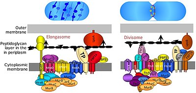
Divisome and elongasome complexes responsible for peptidoglycan synthesis during lateral jail cell-wall growth and sectionalization.[12]
Bacterial jail cell division happens through binary fission or budding. The divisome is a protein complex in bacteria that is responsible for cell division, constriction of inner and outer membranes during division, and peptidoglycan (PG) synthesis at the partitioning site. A tubulin-like protein, FtsZ plays a critical role in formation of a contractile ring for the cell segmentation.[xiii]
Cell Partitioning in Eukaryote [edit]
Cell division in eukaryote is much more complicated than prokaryote. Depending upon chromosomal number reduced or not; Eukaryotic cell divisions can be classified every bit mitosis (equational division) and meiosis (reductional division). A primitive form of cell sectionalization is also institute which is called amitosis. The amitotic or mitotic cell segmentation is more atypical and diverse in the diverse groups of organisms such as protists (namely diatoms, dinoflagellates etc.) and fungi.
-

closed
intranuclear
pleuromitosis -

closed
extranuclear
pleuromitosis -

airtight
orthomitosis -

semiopen
pleuromitosis -

semiopen
orthomitosis -

open
orthomitosis
In mitotic metaphase (meet below), typically the chromosomes (each with 2 sister chromatids that they adult due to replication in the S phase of interphase) arranged and sister chromatids split and distributed towards daughter cells.
In meiosis, typically in Meiosis-I the homologous chromosomes are paired and and then separated and distributed into daughter cells. Meiosis-II is like mitosis where the chromatids are separated. In human and other college animals and many other organisms, the meiosis is chosen gametic meiosis, that is meiosis gives rise to gametes. Whereas in many groups of organisms, especially in plants (observable in lower plants, meiosis just vestigial stage in higher plants), the meiosis gives rise to the kind of spores that germinate into haploid vegetative phase (gametophyte). This kind of meiosis is called sporic meiosis.
Phases of eukaryotic prison cell sectionalisation [edit]
Interphase [edit]
Interphase is the process through which a cell must go before mitosis, meiosis, and cytokinesis.[14] Interphase consists of 3 chief phases: Gi, S, and G2. G1 is a time of growth for the cell where specialized cellular functions occur in lodge to prepare the cell for Deoxyribonucleic acid replication.[15] There are checkpoints during interphase that let the prison cell to either advance or halt farther evolution. One of the checkpoint is between Gane and S, the purpose for this checkpoint is to check for appropriate cell size and any DNA damage . The 2d check bespeak is in the G2 phase, this checkpoint also checks for cell size but also the DNA replication. The concluding check point is located at the site of metaphase, where it checks that the chromosomes are correctly connected to the mitotic spindles.[16] In S phase, the chromosomes are replicated in society for the genetic content to be maintained.[17] During G2, the jail cell undergoes the concluding stages of growth before information technology enters the M phase, where spindles are synthesized. The M phase tin can be either mitosis or meiosis depending on the type of cell. Germ cells, or gametes, undergo meiosis, while somatic cells will undergo mitosis. After the jail cell proceeds successfully through the 1000 phase, information technology may then undergo cell division through cytokinesis. The command of each checkpoint is controlled past cyclin and cyclin-dependent kinases. The progression of interphase is the result of the increased amount of cyclin. As the amount of cyclin increases, more and more than cyclin dependent kinases attach to cyclin signaling the cell further into interphase. At the peak of the cyclin, fastened to the cyclin dependent kinases this system pushes the jail cell out of interphase and into the K phase, where mitosis, meiosis, and cytokinesis occur.[18] There are three transition checkpoints the jail cell has to get through before entering the M phase. The virtually important existence the G1-S transition checkpoint. If the cell does non laissez passer this checkpoint, it results in the jail cell exiting the cell cycle.[19]
Prophase [edit]
Prophase is the first phase of division. The nuclear envelope is broken downwards in this stage, long strands of chromatin condense to class shorter more visible strands called chromosomes, the nucleolus disappears, and microtubules attach to the chromosomes at the disc-shaped kinetochores nowadays in the centromere.[20] Microtubules associated with the alignment and separation of chromosomes are referred to equally the spindle and spindle fibers. Chromosomes will besides be visible under a microscope and volition be connected at the centromere. During this condensation and alignment catamenia in meiosis, the homologous chromosomes undergo a break in their double-stranded Dna at the aforementioned locations, followed by a recombination of the now fragmented parental DNA strands into non-parental combinations, known equally crossing over.[21] This process is evidenced to be caused in a big office past the highly conserved Spo11 poly peptide through a mechanism similar to that seen with toposomerase in DNA replication and transcription.[22]
Metaphase [edit]
In metaphase, the centromeres of the chromosomes convene themselves on the metaphase plate (or equatorial plate), an imaginary line that is at equal distances from the two centrosome poles and held together by complexes known as cohesins. Chromosomes line upward in the middle of the cell by microtubule organizing centers (MTOCs) pushing and pulling on centromeres of both chromatids thereby causing the chromosome to move to the center. At this point the chromosomes are all the same condensing and are currently 1 footstep away from existence the virtually coiled and condensed they volition be, and the spindle fibers have already connected to the kinetochores.[23] During this phase all the microtubules, with the exception of the kinetochores, are in a state of instability promoting their progression towards anaphase.[24] At this indicate, the chromosomes are prepare to carve up into reverse poles of the cell towards the spindle to which they are connected.[25]
Anaphase [edit]
Anaphase is a very brusk stage of the jail cell wheel and it occurs after the chromosomes marshal at the mitotic plate. Kinetochores emit anaphase-inhibition signals until their attachment to the mitotic spindle. Once the final chromosome is properly aligned and fastened the final signal dissipates and triggers the abrupt shift to anaphase.[24] This abrupt shift is acquired by the activation of the anaphase-promoting complex and its function of tagging degradation of proteins important towards the metaphase-anaphase transition. One of these proteins that is broken downwards is securin which through its breakdown releases the enzyme separase that cleaves the cohesin rings holding together the sis chromatids thereby leading to the chromosomes separating.[26] After the chromosomes line upwardly in the heart of the cell, the spindle fibers will pull them autonomously. The chromosomes are split autonomously while the sister chromatids move to opposite sides of the cell.[27] Every bit the sister chromatids are beingness pulled apart, the cell and plasma are elongated by not-kinetochore microtubules.[28]
Telophase [edit]
Telophase is the last phase of the cell wheel in which a cleavage furrow splits the cells cytoplasm (cytokinesis) and chromatin. This occurs through the synthesis of a new nuclear envelope that forms around the chromatin gathered at each pole. The nucleolus reforms every bit the chromatin reverts back to the loose country it possessed during interphase.[29] [30] The sectionalization of the cellular contents is not ever equal and can vary by cell blazon as seen with oocyte formation where 1 of the four daughter cells possess the majority of the cytoplasm.[31]
Cytokinesis [edit]
The terminal stage of the cell division process is cytokinesis. In this stage in that location is a cytoplasmic division that occurs at the end of either mitosis or meiosis. At this phase there is a resulting irreversible separation leading to ii daughter cells. Cell division plays an important role in determining the fate of the cell. This is due to there beingness the possibility of an asymmetric division. This as a effect leads to cytokinesis producing unequal girl cells containing completely different amounts or concentrations of fate-determining molecules.[32]
In animals the cytokinesis ends with formation of a contractile ring and thereafter a cleavage. Simply in plants information technology happen differently. At first a cell plate is formed and then a prison cell wall develops betwixt the 2 daughter cells.
In Fission yeast (S. pombe) the cytokinesis happens in G1 stage [33]
Variants [edit]
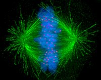
Cells are broadly classified into two main categories: simple not-nucleated prokaryotic cells and circuitous nucleated eukaryotic cells. Due to their structural differences, eukaryotic and prokaryotic cells practice not divide in the same fashion. Also, the pattern of jail cell division that transforms eukaryotic stalk cells into gametes (sperm cells in males or egg cells in females), termed meiosis, is different from that of the division of somatic cells in the body. Image of the mitotic spindle in a human cell showing microtubules in green, chromosomes (Deoxyribonucleic acid) in blueish, and kinetochores in ruddy.
Degradation [edit]
Multicellular organisms replace worn-out cells through cell division. In some animals, all the same, cell division eventually halts. In humans this occurs, on average, after 52 divisions, known as the Hayflick limit. The cell is then referred to as senescent. With each division the cells telomeres, protective sequences of DNA on the end of a chromosome that prevent degradation of the chromosomal Dna, shorten. This shortening has been correlated to negative furnishings such as age related diseases and shortened lifespans in humans.[35] [36] Cancer cells, on the other mitt, are not thought to dethrone in this manner, if at all. An enzyme complex called telomerase, present in big quantities in malignant cells, rebuilds the telomeres through synthesis of telomeric Deoxyribonucleic acid repeats, assuasive division to continue indefinitely.[37]
History [edit]

Kurt Michel with his phase-dissimilarity microscope
A jail cell sectionalisation nether microscope was start discovered past German botanist Hugo von Mohl in 1835 as he worked over the light-green alga Cladophora glomerata.[38]
In 1943, cell division was filmed for the first time[39] past Kurt Michel using a phase-dissimilarity microscope.[40]
Encounter too [edit]
- Binary fission
- Jail cell biology
- Cell fusion
- gametic fusion
- Jail cell growth
- Cyclin-dependent kinase
- Labile cells, cells that constantly divide
References [edit]
- ^ Martin EA, Hine R (2020). A dictionary of biology (6th ed.). Oxford: Oxford University Press. ISBN9780199204625. OCLC 176818780.
- ^ Griffiths AJ (2012). Introduction to genetic analysis (10th ed.). New York: W.H. Freeman and Co. ISBN9781429229432. OCLC 698085201.
- ^ "10.2 The Cell Cycle - Biological science 2e | OpenStax". openstax.org . Retrieved 2020-11-24 .
- ^ Gilbert SF (2000). "Spermatogenesis". Developmental Biology (6th ed.).
- ^ Gilbert SF (2000). "Oogenesis". Developmental Biological science (6th ed.).
- ^ Maton, Anthea (1997). Cells : building blocks of life (tertiary ed.). Upper Saddle River, N.J.: Prentice-Hall. pp. 70–74. ISBN978-0134234762. OCLC 37049921.
- ^ Quammen D (April 2008). "Contagious Cancer". Harper'due south Magazine. ISSN 0017-789X. Retrieved 2019-04-14 .
- ^ Golitsin, Yuri N.; Krylov, Mikhail C. C. (2010). Cell segmentation : theory, variants, and degradation. New York: Nova Scientific discipline Publishers. p. 137. ISBN9781611225938. OCLC 669515286.
- ^ Fletcher, Daniel A.; Mullins, R. Dyche (28 Jan 2010). "Prison cell mechanics and the cytoskeleton". Nature. 463 (7280): 485–492. doi:10.1038/nature08908. ISSN 0028-0836. PMC2851742. PMID 20110992.
- ^ Li, Shanwei; Sun, Tiantian; Ren, Haiyun (27 April 2015). "The functions of the cytoskeleton and associated proteins during mitosis and cytokinesis in plant cells". Frontiers in Plant Science. 6: 282. doi:10.3389/fpls.2015.00282. ISSN 1664-462X. PMC4410512. PMID 25964792.
- ^ Hohmann, Tim; Dehghani, Faramarz (eighteen April 2019). "The Cytoskeleton—A Circuitous Interacting Meshwork". Cells. eight (4): 362. doi:x.3390/cells8040362. ISSN 2073-4409. PMC6523135. PMID 31003495.
- ^ Hugonnet JE, Mengin-Lecreulx D, Monton A, den Blaauwen T, Carbonnelle East, Veckerlé C, et al. (Oct 2016). "Escherichia coli". eLife. 5. doi:x.7554/elife.19469. PMC5089857. PMID 27767957.
- ^ Jail cell Division: The Cycle of the Ring, Lawrence Rothfield and Sheryl Justice, Prison cell, DOI
- ^ Marieb EN (2000). Essentials of human beefcake and physiology (6th ed.). San Francisco: Benjamin Cummings. ISBN978-0805349405. OCLC 41266267.
- ^ Pardee AB (Nov 1989). "G1 events and regulation of cell proliferation". Science. 246 (4930): 603–eight. Bibcode:1989Sci...246..603P. doi:x.1126/science.2683075. PMID 2683075.
- ^ Molinari M (October 2000). "Cell bicycle checkpoints and their inactivation in human being cancer". Jail cell Proliferation. 33 (5): 261–74. doi:x.1046/j.1365-2184.2000.00191.x. PMC6496592. PMID 11063129.
- ^ Morgan DO (2007). The cell wheel : principles of control. London: New Science Press. ISBN9780199206100. OCLC 70173205.
- ^ Lindqvist A, van Zon West, Karlsson Rosenthal C, Wolthuis RM (May 2007). "Cyclin B1-Cdk1 activation continues after centrosome separation to control mitotic progression". PLOS Biology. 5 (5): e123. doi:10.1371/periodical.pbio.0050123. PMC1858714. PMID 17472438.
- ^ Paulovich AG, Toczyski DP, Hartwell LH (February 1997). "When checkpoints fail". Cell. 88 (three): 315–21. doi:10.1016/S0092-8674(00)81870-X. PMID 9039258. S2CID 5530166.
- ^ Schermelleh L, Carlton PM, Haase Due south, Shao 50, Winoto L, Kner P, et al. (June 2008). "Subdiffraction multicolor imaging of the nuclear periphery with 3D structured illumination microscopy". Science. 320 (5881): 1332–6. Bibcode:2008Sci...320.1332S. doi:10.1126/science.1156947. PMC2916659. PMID 18535242.
- ^ Lewontin RC, Miller JH, Gelbart WM, Griffiths AJ (1999). "The Mechanism of Crossing-Over". Modern Genetic Analysis.
- ^ Keeney S (2001). Machinery and control of meiotic recombination initiation. Current Topics in Developmental Biology. Vol. 52. Elsevier. pp. 1–53. doi:10.1016/s0070-2153(01)52008-6. ISBN9780121531522. PMID 11529427.
- ^ "Researchers Shed Light On Shrinking Of Chromosomes". ScienceDaily . Retrieved 2019-04-14 .
- ^ a b Walter P, Roberts Chiliad, Raff K, Lewis J, Johnson A, Alberts B (2002). "Mitosis". Molecular Biology of the Cell (4th ed.).
- ^ Elrod South (2010). Schaum'southward outlines : genetics (5th ed.). New York: Mcgraw-Hill. p. viii. ISBN9780071625036. OCLC 473440643.
- ^ Brooker AS, Berkowitz KM (2014). "The roles of cohesins in mitosis, meiosis, and human health and disease". Cell Cycle Control. Methods in Molecular Biology. Vol. 1170. New York: Springer. pp. 229–66. doi:10.1007/978-1-4939-0888-2_11. ISBN9781493908875. PMC4495907. PMID 24906316.
- ^ "The Cell Cycle". www.biology-pages.info . Retrieved 2019-04-14 .
- ^ Urry LA, Cain ML, Jackson RB, Wasserman SA, Minorsky PV, Reece JB (2014). Campbell Biology in Focus. Boston (Massachusetts): Pearson. ISBN978-0-321-81380-0.
- ^ Dekker J (2014-11-25). "Ii ways to fold the genome during the prison cell cycle: insights obtained with chromosome conformation capture". Epigenetics & Chromatin. 7 (ane): 25. doi:10.1186/1756-8935-seven-25. PMC4247682. PMID 25435919.
- ^ Hetzer MW (March 2010). "The nuclear envelope". Cold Leap Harbor Perspectives in Biology. 2 (3): a000539. doi:10.1101/cshperspect.a000539. PMC2829960. PMID 20300205.
- ^ Gilbert SF (2000). "Oogenesis". Developmental Biology (sixth ed.).
- ^ Guertin DA, Trautmann Southward, McCollum D (June 2002). "Cytokinesis in eukaryotes". Microbiology and Molecular Biology Reviews. 66 (2): 155–78. doi:ten.1128/MMBR.66.2.155-178.2002. PMC120788. PMID 12040122.
- ^ The Cell, Chiliad.M. Cooper; ed ii NCBI bookshelf, The eukaryotic jail cell bike, Effigy fourteen.7
- ^ "Phase Holographic Imaging of Cell Division". Cyberspace archive. Archived from the original on 29 June 2013.
- ^ Jiang H, Schiffer E, Song Z, Wang J, Zürbig P, Thedieck Thou, et al. (August 2008). "Proteins induced by telomere dysfunction and Dna damage represent biomarkers of human being aging and affliction". Proceedings of the National Academy of Sciences of the The states. 105 (32): 11299–304. Bibcode:2008PNAS..10511299J. doi:x.1073/pnas.0801457105. PMC2516278. PMID 18695223.
- ^ Cawthon RM, Smith KR, O'Brien Eastward, Sivatchenko A, Kerber RA (Feb 2003). "Association between telomere length in blood and mortality in people aged sixty years or older". Lancet. 361 (9355): 393–5. doi:10.1016/S0140-6736(03)12384-seven. PMID 12573379. S2CID 38437955.
- ^ Jafri MA, Ansari SA, Alqahtani MH, Shay JW (June 2016). "Roles of telomeres and telomerase in cancer, and advances in telomerase-targeted therapies". Genome Medicine. 8 (1): 69. doi:10.1186/s13073-016-0324-x. PMC4915101. PMID 27323951.
- ^ Biographie, Deutsche. "Mohl, Hugo von - Deutsche Biographie". www.deutsche-biographie.de (in German). Retrieved 2019-04-xv .
- ^ Masters BR (2008-12-15). "History of the Optical Microscope in Cell Biology and Medicine". Encyclopedia of Life Sciences. John Wiley & Sons, Ltd. doi:10.1002/9780470015902.a0003082. ISBN978-0470016176.
- ^ ZEISS Microscopy (2013-06-01), Historic time lapse moving-picture show by Dr. Kurt Michel, Carl Zeiss Jena (ca. 1943), archived from the original on 2021-eleven-07, retrieved 2019-04-15
Further reading [edit]
- Morgan Hi. (2007). "The Jail cell Cycle: Principles of Control" London: New Scientific discipline Press.
- J.K.Turner Fetus into Man (1978, 1989). Harvard University Printing. ISBN 0-674-30692-9
- Prison cell partitioning: binary fission and mitosis
- McDougal, W. Scott, et al. Campbell-Walsh Urology Eleventh Edition Review. Elsevier, 2016.
- The Mitosis and Prison cell Bicycle Control Section from the Landmark Papers in Cell Biology (Gall JG, McIntosh JR, eds.) contains commentaries on and links to seminal research papers on mitosis and jail cell division. Published online in the Paradigm & Video Library of The American Society for Cell Biology
- The Image & Video Library Archived 2011-06-10 at the Wayback Automobile of The American Order for Cell Biological science contains many videos showing the prison cell division.
- The Cell Segmentation of the Prison cell Prototype Library
- Images : Calanthe discolor Lindl. - Flavon's Secret Flower Garden
- Tyson's model of jail cell division and a Description on BioModels Database
- WormWeb.org: Interactive Visualization of the C. elegans Cell Lineage - Visualize the unabridged set of jail cell divisions of the nematode C. elegans
Source: https://en.wikipedia.org/wiki/Cell_division
0 Response to "what term is used to describe new cells"
Post a Comment